Envision Eye Hospital

Welcome to Envision Eye Hospital, a Tertiary Care Superspeciality Eye Hospital offering complete Eye Care under one roof, ranging from complex Retinal laser surgeries, LASIK laser correction of glass numbers to simple and complicated cataract procedures, using advanced technology and updated surgical equipment. It has always been our endeavor to offer high standards of quality eye care at very affordable costs.

From the most simple to the most complex eye disease to providing eye care from infancy up to old age, we have expert ophthalmologists and specialists to take care of our patients. With many firsts to its credit, Envision Eye Hospital has always brought in the latest and the best tech under its fold to benefit one and all.

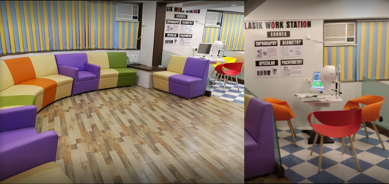
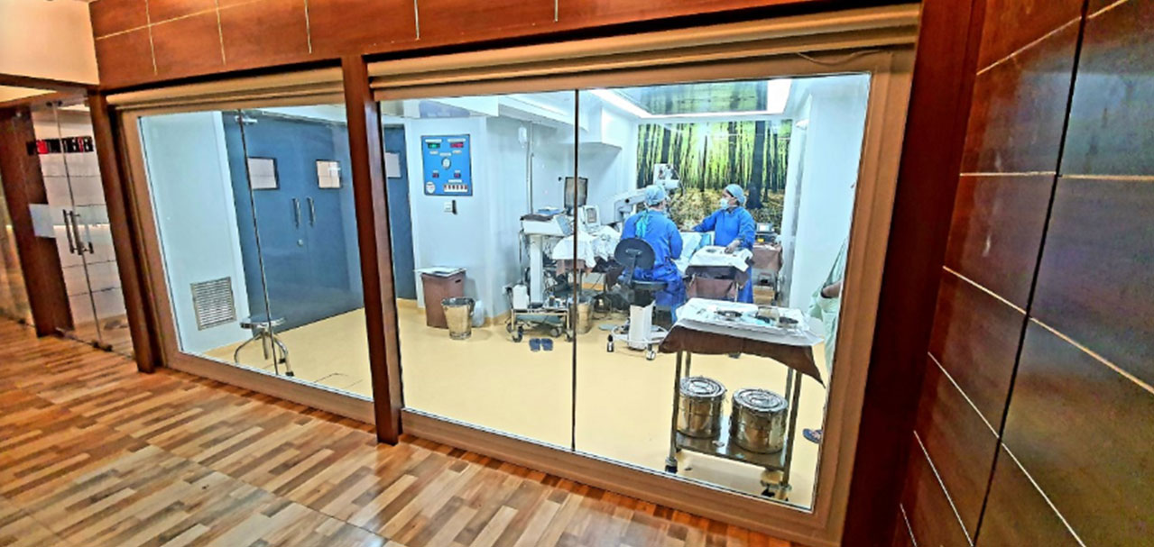
Our NABH-accredited world-class ultra-modern Eye Care facility spanning 3 floors and spread over 5000 sq ft. is strategically located on S V Rd, the arterial central road of the western suburbs, 2 mins walking distance from the Jogeshwari railway station and 5 mins driving distance from the western express highway.

WITH AN ADDITIONAL PROVISION OF AN

  • In-Hospital Pathology laboratory that caters to pre-operative blood investigations and anesthesia fitness so that the patient does not have to wander around to get medical tests done prior to surgery.
  • Indoor admission facility with private and semi-private rooms for long-distance or overseas patients.
  • Intensive care unit, round-the-clock doctors and nursing staff as well as a dedicated ophthalmology pharmacy in the same premises on the ground floor (Obaid Medical Stores).

Envision eye hospital stands a step ahead in handling eye emergencies, eye trauma cases, and eye cases with critical systemic conditions.


VISION STATEMENT

To become the highest level super specialty ophthalmology and retina center providing international standards at affordable rates.

MISSION STATEMENT

The mission of the Hospital is to continuously strive to achieve excellence in eye care and provide services with cutting-edge technology, compassion, and complete transparency.

VALUE STATEMENT

The working of EEH is entirely ethical and transparent, directed towards affordable healthcare, without any discrimination of caste, race, religion, or creed.

QUALITY POLICY

We shall strive to achieve a leadership position in comprehensive Eye Care by providing the most effective equipment, trained manpower & infection-free environment while meeting the needs and expectations of patients & society at large.

We commit to being an organization that:

  • Meets all the regulatory & obligatory requirements.
  • Promotes respect & protects patient's rights, responsibilities, and education
  • Implement "Entry Level NABH Standard for SHCO" in true spirit.
  • Adheres to the best medical & infection control practices.
  • Encourages feedback to enhance patients' satisfaction


OUR ADVANCED SURGICAL EQUIPMENT

LASIK MACHINE [SHWIND]

BLADELESS, TOUCHLESS, FLAPLESS : THE WORLD'S 1ST LASIK SUITE (Freedom from Glasses)

CUSTOM OR CONTOUR GUIDED LASIK : In accordance with one's aberration profile to ensure Hi-definition Vision.

BLADELESS, TOUCHLESS, FLAPLESS : World's 1st PATENTED SMART SURF SMART PULSE TECHNOLOGY (SPT) Trans-PRK LASIK.

PRECISE : Laser with the Smallest Beam Profile (0.5 mm) in the world for the most accurate results and maximum tissue preservation.

FAST : 500 HZ tissue ablation (1 D in 2 seconds) speed ensures quick treatment with excellent co-operation in anxious patients.

SAFE : Laser with 1he 6-DIMENSIONAL FASTEST EYE TRACKER (1050 HZ) monitors eye movements with the quickest reaction time.

PAINLESS : Patented technologies such as AUTOMATIC FLUENCE LEVEL ADJUSTMENT (AFLA), INTELLIGENT THERMAL EFFECT CONTROL (ITEC) ensures rapid cooling between laser pulses.

PRESBYOPIC LASIK : LASIK possible AT AGE 40 AND BEYOND AND EVEN AFTER CATARACT SURGERY

DORC VITRECTOMY MACHINE

The DORC vitrectomy system has been designed with the highest cut rate available and utilizes an enhanced fluidics system for vacuum or flow vitrectomy modes.

ROBOT PHACO AND VITRECTOMY MACHINE

The machine delivers an exceptional level of performance through its advanced technologies, allowing surgeons to treat eye conditions that may have once been considered untreatable. Recognized as the one of world’s most advanced machines for 23G 25G and 27G vitreoretinal surgery, the ROBOT system combines advancements in high-speed cutting, intraocular pressure control, illumination, laser and other features allowing for more surgeon control during retinal surgery. The ROBOT system has combined capabilities to perform advanced sutureless vitrectomy/retinal surgery and retinal lasers with endo-laser facility for diabetic retinopathy and managing complicated retinal detachment cases.

ZEISS 160 SURGICAL MICROSCOPE

As Multifaceted as the Challenges of Ophthalmic Surgery OPMI VISU® 160 was designed specifically for hospitals that perform a broad spectrum of ophthalmic surgical procedures. The versatile configuration and accessory options are tailored to meet the needs of the surgeon:

  • Deep View function, the depth of field management system that allows you to choose between optimized depth perception or maximum light transmission.
  • Integrated slit illumination for true retro-illumination during lens extraction.
  • Ergonomic handgrips for precise positioning.
  • Apochromatically corrected optics.
  • With BrightFlex illumination, consisting of 2° and fading 6° illumination, for a clear red reflex and good light conditions.
  • Extensive array of accessory options, such as co-observation, video and digital recording/editing systems.

Wide angle viewing system, BIOM, OCULUS (Germany)

Oculus BIOM system for non-contact high resolution and wide field retinal surgery

Endolaser, VISULAS 532, ZEISS (Germany)

Used in retinal surgeries to laser retinal tears and leakage areas in the retina

Live surgery recording system, SONY (Japan)

Allows the patient’s relatives to view the surgery being performed live and also provide the patients recording of their own surgery

Class-B vacuum autoclave, HANSEN (Korea)

Compact, high performance autoclave using vacuum pump to sterilize surgical instruments

ETO sterilizer, RUJIKON (India)

For sterilizing surgical instruments by chemical method to ensure safe and sterile process

OUR ADVANCED DIAGNOSTIC EQUIPMENT

Angiography OCT

This non-invasive dye free technique as against traditional invasive dye technique allows the visualization of the microvasculature of the retina

Read More.. Read less

without the risk of injections and dye related allergic side-effects. Angiography scan allows assessment of the structural vasculature of the macula, the periphery or the optic disc with extremely short scanning times of 1,6 seconds in standard resolution or 3 seconds in high resolution.

Fundus Camera with autofluorescence, VISUCAM 500, Zeiss (Germany)

For capturing fundus (retina) photograph The main

structures that can be visualised are the central and peripheral retina, optic disc and macula

FUNDUS FLUORESCEIN ANGIOGRAPHY

A fluorescein angiogram looks at the blood vessels and the lining at the back of your eye (the retina). This test can pick up changes in the condition

Read More.. Read less

of your eyes. It can help the specialist make a diagnosis or tell them how well treatment is working.

Indocyanine green angiography, VISUCAM 500, Zeiss (Germany)

Apart from fluorescein dye, our machine is also

equipped to perform indocyanine green angiography to detect deeper leaks of the choroid

Corneal topographer and dual-schiemflug camera, SIRIUS, CSO (Italy)

Takes corneal curvature of anterior and posterior Surfaces long with aberration measurements,

Read More.. Read less

a pre-requisite before LASIK surgery and premium lens implant surgery.

Corneal pachymeter
SIRIUS, CSO (Italy)

It is the process of measuring corneal thickness.

It is performed prior to lasik and for keratoconus screening

Specular Microscope
NIDEK (Japan)

Measures corneal endothelial counts, a pre-requisite for cataract surgery. This device is available at very few selected centres in mumbai

A-scan and optical biometer, OA-2000 TOMEY (JAPAN)

Takes accurate lens measurements calculates

Cylindrical axis which defines accurate incision & lens placement

Ultrasound A-scan, AXIS-II, QUANTEL(France)

To measure the length of the eye and calculate the lens power before cataract surgery

Blue on yellow perimetry (SWAP), HUMPHREY 745i, ZEISS (Germany)

Blue on yellow perimetry detects glaucoma even before it is detected symptomatically

White on white perimetry (HVF)

Humphrey field analyser (HFA) is a tool for measuring the human visual field

Read More.. Read less

,particularly for detecting monocular visual field. The results of the analyser identify the type of vision defect. Therefore, it provides information regarding the location of any disease processes or lesion(s) throughout the visual pathway.

YAG Laser, Appasamy Associates

Laser for removing membrane behind the lens

(after cataract) and treatment of various types of glaucoma etc.

Optical Coherence Tomography, 3D SPECTRAL DOMAIN CIRRUS, Zeiss

Takes high resolution cross-sections of the cornea and lens to detect deeper pathologies.

Meibography

Meibography and Tear- break up time (TBUT) SIRIUS, CSO (ITALY) Helps in detailed evaluation in dry eye and computer vision syndrome

BSCAN

B-Scan Ocular Ultrasound Devices / B Scan of the Eye B-scan Ultrasonography, often called just

Read More.. Read less

B-scan or Brightness scan, offers two-dimensional cross-sectional view of the eye as well as the orbit. A B-scan is used on the outside of the closed eyelid to view the eye. This type of diagnostic tool is most helpful when there is difficulty examining the eye normally. A B-scan can help accurately view other eye structures like the lens, choroid, sclera, vitreous and retina. A B-scan is very helpful in diagnosing retinal detachment.

Gonioscope, Volk (Germany)

Special lens which helps in visualising the anterior chamber angle for diagnosing various types of glaucoma

OUR BASIC OPHTHALMIC EVALUATION EQUIPMENT

Non-Contact Tonometer’s / Air-Puff Tonometer

A tonometer is a diagnostic tool used by eye care professionals to

Read More.. Read less

measure the intraocular pressure (IOP) inside a patient's eyes. A patient's IOP can help an ophthalmologist or optometrist determine a patient's risk for developing glaucoma — a disease that damages the optic nerve of the eye, usually resulting in vision loss or even blindness. A non-contact tonometer uses a small puff of air (which is why its many times referred to as the "puff test") to measure an eye's pressure. Known as pneumotonometry, the air puff flattens the patient's cornea in a non-invasive way, meaning eyes do not need to be numbed prior to the test.

Slit-lamp with live slit lamp photography, APPASAMY (India)

For examination of the eye. Slit lamp photography for demonstration of the findings and capturing the images

Indirect ophthalmoscopy,
KEELER (UK)

For examination of the retina and diagnosing retinal diseases

Fully automated autorefractometer with phoropter

A phoropter is a computer-controlled machine used during an eye examination to provide

Read More.. Read less

an objective measurement of a person's refractive error and prescription for glasses or contact lenses. This is achieved by measuring how light is changed as it enters a person's eye.

Applanation tonometry
APPASAMY (India)

Gold standard for intraocular pressure measurement

LED vision chart
AUROLAB (India)

For testing visual acuity, contrast sensitivity and colour blindness

Autolensometer, Autorefractometer/ Autokeratometer, NIDEK (Japan)

Accurate measurement of spectacle numbers